Breasts are an essential anatomical and functional part of the human body, present in both People of all genders. While male breasts remain undeveloped throughout life, female breasts undergo significant changes, particularly during puberty, pregnancy and lactation. Understanding the structure and function of the breast is important for students of anatomy, medical science and anyone interested in human biology.
What are breasts?
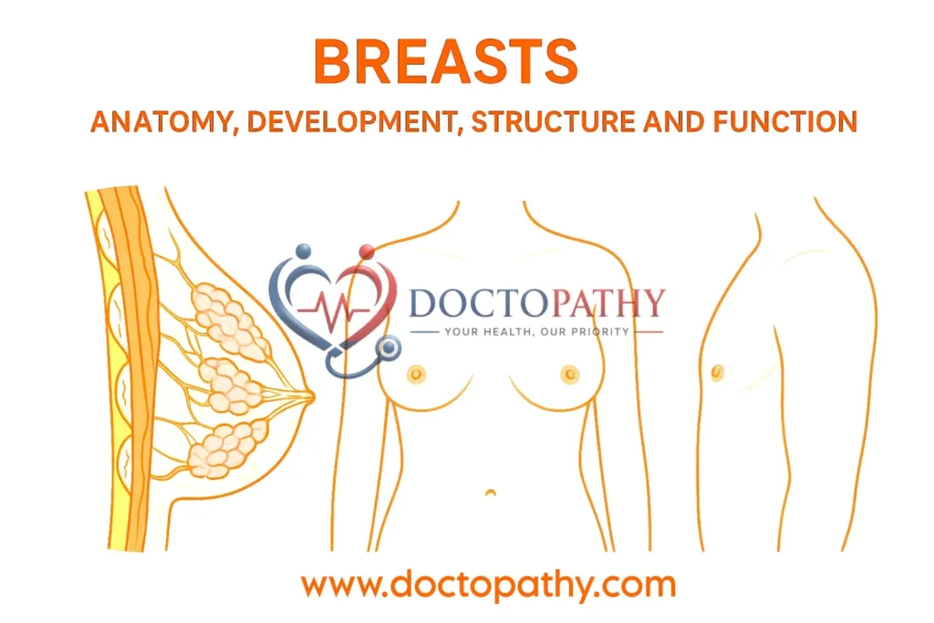
The breasts (mammary glands) are specialized organs located on the chest. They are composed of glandular tissue, fibrous tissue and fatty tissue. Their primary biological function is to produce and secrete milk during lactation. Both People of all genders have breasts, but: In men the breasts remain rudimentary. In women they grow and develop during puberty and especially during pregnancy and breastfeeding.
Female breast anatomy
1. Location and size
In young women, the breasts are hemispherical structures located on the anterior thoracic wall . They extend
vertically from the second to the sixth ribs and horizontally from the lateral border of the sternum to the mid-axillary line.
Axial tail
A superolateral extension called the axillary tail of Spence runs into the armpit region and is closely associated with the axillary lymph nodes .
2. Retromammary space
A loose areolar space behind the breast allows it to glide freely over chest muscles such as:
- Major pectoralis major
- serratus anterior
- external oblique
This space separates the breast from the deep fascia.
External features of the breast
1. Nipple
The nipple is a small, conical protuberance usually located in the fourth intercostal space. These include:
- 15–20 lactiferous duct openings
- Smooth muscle fibers that cause the nipple to twitch or contract
Retraction of the nipple during development can cause difficulty in breastfeeding.
2. Areola
The pigmented area around the nipple is called the areola. Changes in color:
- Pink/light in virgins
- Darkening during pregnancy
- Gets lighter after breastfeeding
The areolar glands enlarge during pregnancy and secrete an oily substance that protects the skin during breastfeeding.
internal structure of the breast
The breast is made up of three main components:
1. Glandular tissue
- It has 15-20 lobes.
- The lobes consist of smaller lobules
- Lobules contain alveoli (milk-secreting structures)
Each lobe drains into a lactiferous duct, which widens into a lactiferous sinus – a temporary milk reservoir.
2. Fibrous tissue
Connective tissue forms the Cooper ligaments, which support the breast.
In breast cancer, these ligaments can shrink, causing dimpling of the skin .
3. Fatty tissue
It surrounds the glandular tissue and largely determines the size and shape of the breast.
Breast development (mammogenesis)
Before puberty
- Only ducts are present
- No alveoli
- Growth primarily due to fat accumulation
On youth
Estrogen stimulates:
- Branching of the ducts
- Formation of terminal buds
- Development of the initial alveoli
During pregnancy
Hormones involved: estrogen, progesterone, prolactin, growth hormone
The changes include:
- Formation of true milk-secreting alveoli
- Increased duct branching
- Production of colostrum (yellowish, nutrient-rich fluid)
Lactation
It begins 4-5 days after the baby is born. Stimulated by the baby’s suckling, the myoepithelial cells contract to push milk through the ducts.
After breastfeeding
- Milk stops
- alveoli shrink
- Glandular tissue decreases
After menopause
- Glandular tissue atrophy
- Breasts become smaller and thicker
Male breasts
Male breasts remain underdeveloped, including:
- Basic ducts (fewer in number)
- some fibrous tissue
- thick
These usually do not develop functional alveoli.
Blood supply, lymphatic drainage and nerves
Blood supply
Where do breasts get blood from?
- Axillary artery branches
- internal thoracic artery
- intercostal arteries
Venous drainage
The veins form a network around the nipple called the venous circle, which drains into the:
- axillary vein
- internal thoracic vein
Nerve supply
From the 4th-6th thoracic nerves, providing:
- Sensory Supplies
- Sympathetic Fibers
Hormonal control, not nerves, is responsible for milk secretion.
Conclusion
Breasts are complex organs with significant structural, functional and hormonal significance, particularly in women. From puberty to pregnancy and menopause, breast tissue undergoes significant hormonally controlled changes. Understanding the anatomy and physiology of the breasts is essential for medical students, healthcare professionals and educational content creators.